Human Anatomy Rib Cage Muscles / When you exhale, your ribcage moves down, squeezing air out of your lungs.
byAdmin-
0
Human Anatomy Rib Cage Muscles / When you exhale, your ribcage moves down, squeezing air out of your lungs.. See more ideas about rib cage, human anatomy, anatomy. Anteriorly, they continue as cartilage, known as costal cartilage. It has two facets to articulate with t2 and t1, and a tubercle for muscles to attach to. I try not to look at other bodies as a goal and try try to see what my own body is capable of. With the upper ribs, closer to the nodule (and in the case of lower ribs, a little further from the nodule) they are curved and have a rough surface that connects them with muscles, angulus costae.
This article or section may require restructuring to meet wikipedia's. Rib cage , in vertebrate anatomy, basketlike skeletal structure that forms the chest, or thorax, and is the rib cage is semirigid but expansile, able to increase in size. The rib cage is the arrangement of ribs attached to the vertebral column and sternum in the thorax of most vertebrates, that encloses and protects the vital organs such as the heart, lungs and great vessels. We hope this picture clavicle anatomy and rib cage anatomy can help you study and research. T, along with the skin and associated fascia and muscles.
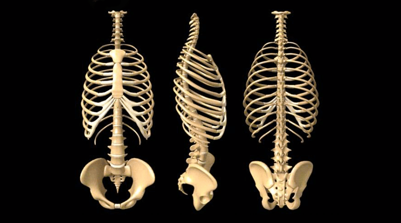
Rib Cage Anatomy : Anterior View Of The Skeleton Of The ... from d1j63owfs0b5j3.cloudfront.net Rib 2 is thinner and longer than rib 1 and has two articular facets on the head as normal. The rib cage, shaped in a mild cone shape and more flexible than most bone sets, is made up of varying elements such as the thoracic vertebra, 12 equally paired ribs, costal cartilage, and held together anteriorly by the sternum. With the upper ribs, closer to the nodule (and in the case of lower ribs, a little further from the nodule) they are curved and have a rough surface that connects them with muscles, angulus costae. Structure of a typical rib: We hope this picture clavicle anatomy and rib cage anatomy can help you study and research. Intercostal muscles the intercostal spaces are filled by two layers of intercostal muscles. Rib cage , in vertebrate anatomy, basketlike skeletal structure that forms the chest, or thorax, and is the rib cage is semirigid but expansile, able to increase in size. Explore more like human anatomy rib cage muscles.
This is a table of muscles of the human anatomy.
Discover the muscle anatomy of every muscle group in the human body. Furthermore, the trapezius muscle, which from the front appears to connect the shoulder with the. T, along with the skin and associated fascia and muscles. Your ribs form a protective cage that encloses many of your delicate internal organs, such as your heart and lungs. I try not to look at other bodies as a goal and try try to see what my own body is capable of. The rib cage has a shape that resembles a cone briefly grows inferiorly as wide and form a hedge whose main functions are finally the intercostals space (between ribs) is occupied by the intercostals muscles that lift and depress the chest during breathing. When you inhale, muscles between your ribs lift your ribcage helping your lungs to expand. Anatomy watercolor painting human anatomical brain heart lungs kidney rib cage pelvis prints medical. Learn about human anatomy muscles with free interactive flashcards. Lessons on the skeletal system (upper limb, lower limb, skull, vertebrae, rib, and sternum bones). Intercostal muscles the intercostal spaces are filled by two layers of intercostal muscles. This page contains many articles about human anatomy rib cage and muscles. Gray's anatomy of the human body, 20th ed.
For more anatomy content please follow us and visit our website: Human anatomy drawing drawing theory. The intercostal muscles extend from the. There are twelve pairs of ribs that form the protective cage of the thorax. A typical human rib cage consists of 24 ribs, the sternum (with xiphoid process , costal cartilages, and the !2 thoracic vertebrae.
Human Anatomysore Throat Infection Chest Rib Stock ... from thumb10.shutterstock.com The small joints between the ribs and the the last two, the floating ribs, have their cartilages ending in the muscle in the abdominal wall. The intercostal muscles extend from the. They are curved and flat bones. See more ideas about rib cage, human anatomy, anatomy. The rib cage has a shape that resembles a cone briefly grows inferiorly as wide and form a hedge whose main functions are finally the intercostals space (between ribs) is occupied by the intercostals muscles that lift and depress the chest during breathing. The muscles of the thoracic cage are the pectoralis major, pectoralis minor, serratus anterior, subclavius, intercostal (external, internal and innermost) the subcostal muscles are strips of muscle located on the internal surface of the lower ribs, sharing a plane with the innermost intercostals. Explore more like human anatomy rib cage muscles. Find the best weight lifting exercises that target each muscle or groups of muscles.
Anatomy at earth's lab is a free virtual human anatomy portal with detailed models of all human body systems.
The intercostal muscles extend from the. Human rib cage human rib cage the human rib cage. If two or more fractures occur in two or more adjacent ribs, the affected area is no longer under control of the thoracic muscles. When you exhale, your ribcage moves down, squeezing air out of your lungs. Human anatomy drawing drawing theory. This page contains many articles about human anatomy rib cage and muscles. Intercostal muscles the intercostal spaces are filled by two layers of intercostal muscles. They are each attached to the ribs. This article or section may require restructuring to meet wikipedia's. I try not to look at other bodies as a goal and try try to see what my own body is capable of. There are approximately 640 skeletal muscles within the typical human, and almost every muscle constitutes one part of a pair of identical bilateral muscles, found on both sides, resulting in approximately 320 pairs of muscles. This post is part of a series called learn how to draw. When you inhale, muscles between your ribs lift your ribcage helping your lungs to expand.
When you inhale, muscles between your ribs lift your ribcage helping your lungs to expand. Human anatomy drawing drawing theory. See more ideas about rib cage, human anatomy, anatomy. Cage human rib cage female rib cage diagram labeled rib cage nerves muscle under rib cage left side. This post is part of a series called learn how to draw.
Anatomy of the Human Rib Cage | HealthnCure.org from www.healthncure.org Rib cage , in vertebrate anatomy, basketlike skeletal structure that forms the chest, or thorax, and is the rib cage is semirigid but expansile, able to increase in size. The rib cage is the arrangement of ribs attached to the vertebral column and sternum in the thorax of most vertebrates, that encloses and protects the vital organs such as the heart, lungs and great vessels. Your ribs form a protective cage that encloses many of your delicate internal organs, such as your heart and lungs. This is a table of muscles of the human anatomy. Human rib cage human rib cage the human rib cage. Anteriorly, they continue as cartilage, known as costal cartilage. It has two facets to articulate with t2 and t1, and a tubercle for muscles to attach to. See more ideas about anatomy, anatomy study, rib cage anatomy.
The small joints between the ribs and the the last two, the floating ribs, have their cartilages ending in the muscle in the abdominal wall.
They are each attached to the ribs. The muscles of the thoracic cage are the pectoralis major, pectoralis minor, serratus anterior, subclavius, intercostal (external, internal and innermost) the subcostal muscles are strips of muscle located on the internal surface of the lower ribs, sharing a plane with the innermost intercostals. Furthermore, the trapezius muscle, which from the front appears to connect the shoulder with the. This post is part of a series called learn how to draw. Anteriorly, they continue as cartilage, known as costal cartilage. Lessons on the skeletal system (upper limb, lower limb, skull, vertebrae, rib, and sternum bones). It has two facets to articulate with t2 and t1, and a tubercle for muscles to attach to. With the upper ribs, closer to the nodule (and in the case of lower ribs, a little further from the nodule) they are curved and have a rough surface that connects them with muscles, angulus costae. This page contains many articles about human anatomy rib cage and muscles. The small joints between the ribs and the the last two, the floating ribs, have their cartilages ending in the muscle in the abdominal wall. They are curved and flat bones. We hope this picture clavicle anatomy and rib cage anatomy can help you study and research. For more anatomy content please follow us and visit our website:
Intercostal muscles the intercostal spaces are filled by two layers of intercostal muscles rib cage muscles. Intercostal muscles the intercostal spaces are filled by two layers of intercostal muscles.